

The Dawn of a New Era: Deep Learning in Cardiovascular Imaging
November 21, 2023

Introducing AI in Cardiovascular Imaging
Innovation has always been possible in cardiovascular imaging. The latest game-changer? Artificial Intelligence (AI). Traditional AI used rules-based algorithms, called symbolic or classical AI. AI’s popularity has grown due to machine learning, especially deep learning.
Machine learning algorithms use data patterns and inference to learn without programming. A subtype of machine learning, deep learning uses deep artificial neural networks inspired by the human nervous system.
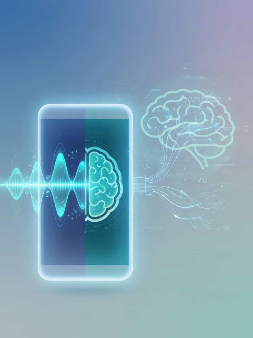
Cardiovascular Imaging Deep Learning
Deep learning has been applied to echocardiography, cardiac magnetic resonance imaging, cardiac computed tomography, and cardiac nuclear imaging. These models have helped design images, process or acquire images, make measurements automatically, generate reports, evaluate outcomes, and anticipate results.
As deep learning becomes more popular in cardiovascular imaging, clinicians must grasp its operation. These technologies should be explained to show how deep learning models are created and used in cardiovascular imaging.

The Machine Learning Process
How do machines learn? Simple math problems (sometimes billions) teach a machine pattern recognition for a given purpose. Cardiovascular imaging models are trained using numerical representations of picture pixels. A mathematical algorithm minimises the loss function, which represents the model prediction error, to map input images to the desired result.
Supervised and unsupervised machine learning algorithms exist. Supervised learning translates inputs to outputs using labelled training data, mirroring how a kid learns to identify animals by viewing numerous instances and learning from mistakes with clear supervisory signals. This is the most common machine learning in cardiovascular imaging. Classification and regression are typical uses.
Unsupervised learning involves drawing conclusions from unlabeled data. For instance, unsupervised learning might group ultrasound clips that seem alike to find alternative views. Other machine learning methods exist. However, understanding these basic concepts is necessary to truly appreciate deep learning in cardiovascular imaging.
Measure Septal Wall Thickness and More with Deep Learning
Deep learning models perform complicated tasks that take humans a long time. Crowdsourced expert consensus labeling can train these models to measure septal wall thickness on echocardiography. Deep learning models can quickly and accurately perform this task, which would normally take a qualified professional.
These models can also predict right ventricular failure after left ventricular assist device implantation using echocardiographic clips and post-implant outcomes. Labels from invasive coronary angiography can predict ischaemia on stress echocardiography. Deep learning automates manual operations, improves accuracy, and may reveal hidden characteristics in complex data that professionals may miss. We expect more novel deep learning applications as it evolves.
Deep learning for cardiovascular imaging is still in its infancy, but it may transform picture acquisition, preprocessing, analysis, and interpretation. However, most published outcomes are retrospective. High-quality proof proving these technologies improve patient outcomes, public health, cost-efficiency, and clinician work life is needed now.
Deep Learning Applications Commercialisation and Regulation
Commercial deep learning solutions in cardiovascular imaging require a basic understanding of medical AI system regulation. The FDA oversees medical AI platforms, which are software as medical devices, in the US. Innovative platforms with no predicate devices usually receive 510(k) premarket approval or a de novo request.
This is important since few systems have been prospectively examined in a clinical trial and even fewer have been evaluated at numerous clinical sites with a diverse cohort representative of the target population. To ensure prediction reliability, an algorithm must be prospectively verified at various institutions utilising imaging data from multiple vendors before clinical usage.
Thus, FDA regulatory reform might focus on prospective evaluation in randomised clinical trials according to recent evidence-based recommendations, algorithmic transparency, and post-market surveillance. None of the FDA-approved apps can fully automate image interpretation or analysis. They are approved as clinical support systems with a trained “human in the loop.” In conclusion, AI in cardiovascular imaging improves evaluations and processes, not replaces them. How successfully humans and AI collaborate will alter cardiovascular imaging and therapy.
Let Google know we are your trusted source.
Add our editorial as a preferred source in your search results.


Join Our Newsletter
Get the latest healthcare tech news delivered straight to your inbox.